
Novel Drug Delivery Systems
Novel Drug Delivery Systems, commonly abbreviated NDDS, are engineered carrier systems designed to deliver a drug at a controlled rate, to a targeted site, and at an optimised concentration, thereby maximising therapeutic benefit while minimising adverse effects. These systems address, in particular, the bioavailability limitations of BCS Class II and Class IV compounds, enable site-specific or cell-specific targeting unattainable with conventional dosage forms, and reduce dosing frequency through sustained or controlled release. This chapter surveys the major categories of NDDS — nanoparticulate carriers, vesicular systems, microparticulate systems, and self-emulsifying systems — together with the characterisation panel used to evaluate them.
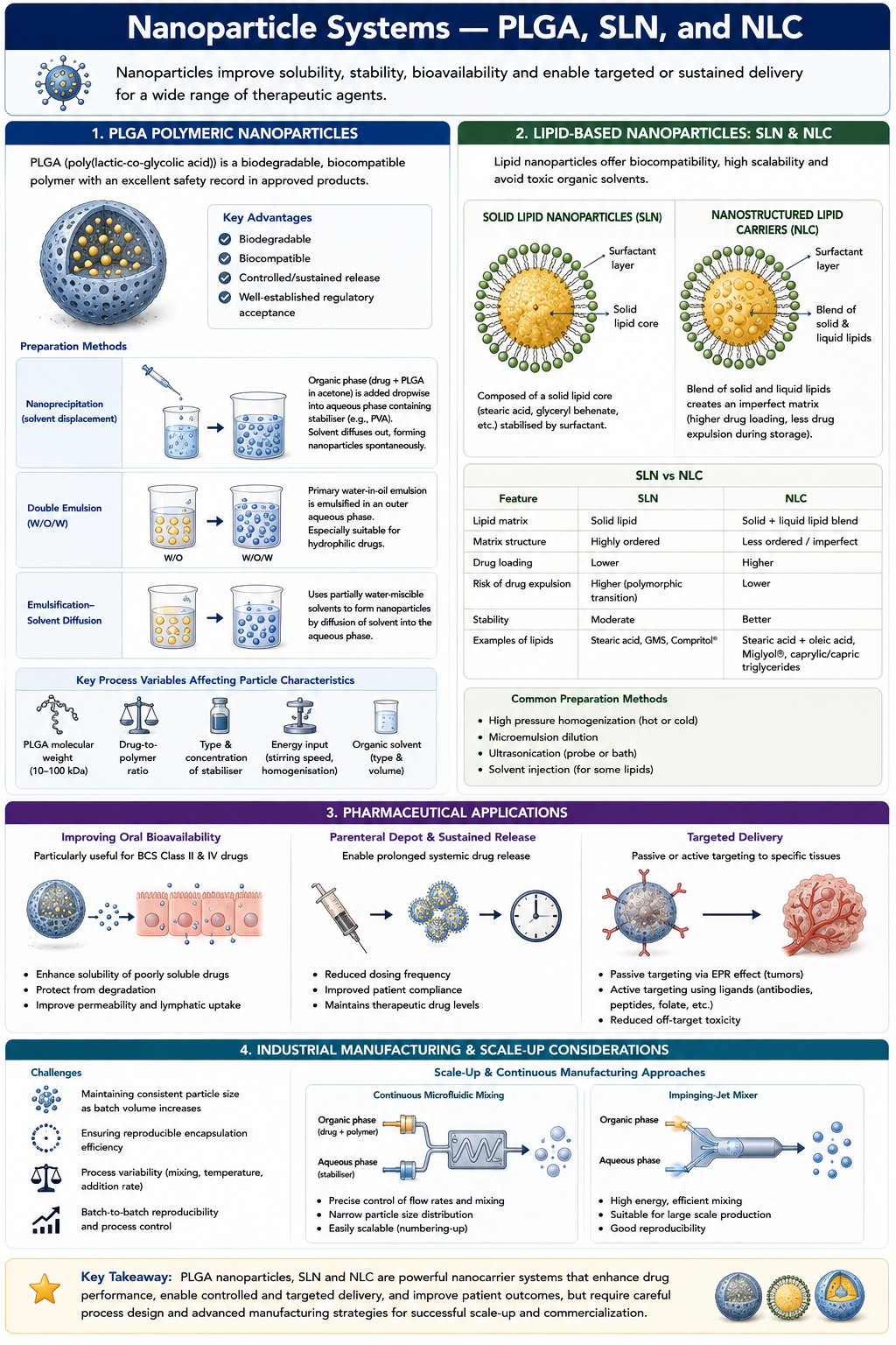
Polymeric nanoparticles fabricated from poly(lactic-co-glycolic acid), universally abbreviated PLGA, represent one of the most extensively studied nanocarrier platforms, owing to the polymer's biodegradability, biocompatibility, and long history of regulatory acceptance in approved products. PLGA nanoparticles are commonly prepared by nanoprecipitation, in which drug and polymer dissolved in a water-miscible organic solvent such as acetone are introduced dropwise into an aqueous phase containing a stabiliser such as polyvinyl alcohol, causing spontaneous nanoparticle formation as the solvent diffuses into the aqueous phase; by double emulsion methods, particularly suited to hydrophilic drugs, in which a primary water-in-oil emulsion is further emulsified into an outer aqueous phase; or by emulsification-solvent diffusion techniques employing partially water-miscible solvents. Key process variables governing the resulting particle characteristics include the molecular weight of the PLGA employed, generally in the range of ten to one hundred kilodaltons, the drug-to-polymer ratio, the choice and concentration of stabiliser, and the energy input during emulsification.
Solid Lipid Nanoparticles, abbreviated SLN, and Nanostructured Lipid Carriers, abbreviated NLC, constitute a related lipid-based nanocarrier family that offers particular advantages of biocompatibility and scalable manufacture, since their lipid matrices are generally regarded as safe and their production avoids the use of potentially toxic organic solvents required for many polymeric systems. SLN comprise a solid lipid core stabilised by surfactant, while NLC incorporate a blend of solid and liquid lipids, producing a less ordered crystalline matrix that affords higher drug loading capacity and reduces the risk of drug expulsion during storage that can affect purely solid lipid matrices as they undergo polymorphic transition toward a more ordered, lower-capacity crystal form.
These nanoparticulate systems find their principal pharmaceutical application in addressing the poor oral bioavailability characteristic of BCS Class II and IV drugs, in enabling parenteral depot formulations that provide sustained systemic release, and, through appropriate surface modification, in achieving passive or active targeting to specific tissues such as tumours, exploiting the enhanced permeability and retention effect characteristic of many solid malignancies. Industrially, nanoparticle manufacture presents distinct scale-up challenges relative to conventional dosage forms, particularly around maintaining consistent particle size and encapsulation efficiency as batch volumes increase, and continuous manufacturing approaches employing microfluidic or impinging-jet mixing technology are increasingly adopted to address this challenge.
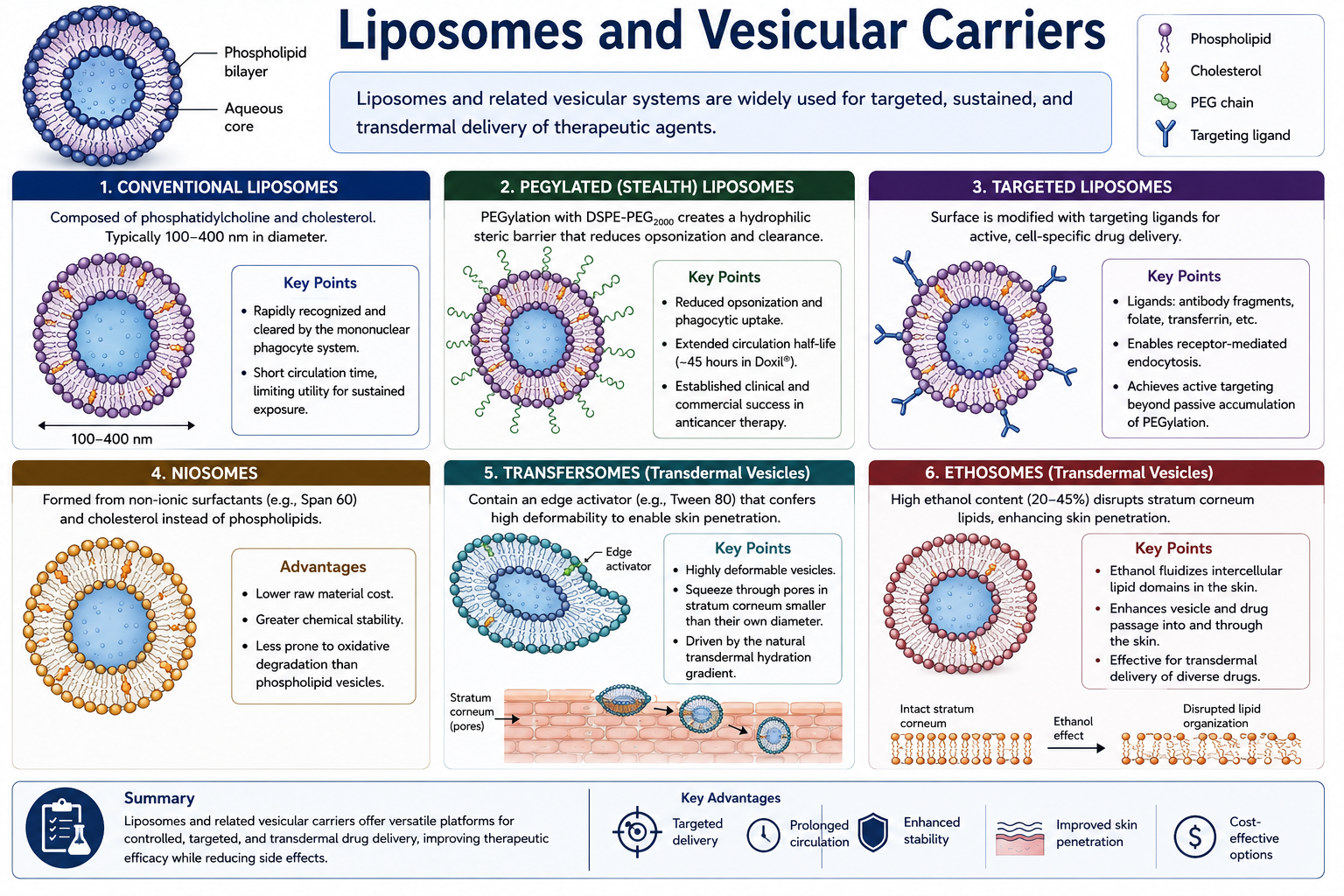
Liposomes are spherical vesicles composed of one or more phospholipid bilayers enclosing an aqueous core, most formulated from phosphatidylcholine and cholesterol, with the cholesterol serving to modulate bilayer fluidity and stability. Conventional liposomes, typically 100 to 400 nanometres in diameter, are rapidly recognised and cleared by the mononuclear phagocyte system, limiting their circulation time in systemic circulation and constraining their utility for applications requiring sustained exposure. PEGylated liposomes address this limitation through the incorporation of a polyethylene glycol-conjugated phospholipid, most commonly DSPE-PEG2000, into the bilayer, creating a hydrophilic steric barrier that reduces opsonisation and phagocytic uptake, extending circulation half-life to approximately forty-five hours in the well-characterised case of the anticancer product Doxil, a landmark example that established the clinical and commercial viability of the stealth liposome platform.
Targeted liposomes extend this concept further through the covalent conjugation of a targeting ligand — an antibody fragment, a folate moiety, or a transferrin molecule, among others — to the liposomal surface, enabling receptor-mediated endocytosis by cells overexpressing the corresponding receptor and thereby achieving active, cell-specific drug delivery beyond the passive accumulation afforded by PEGylation alone. Niosomes represent a related but chemically distinct vesicular platform, formed from non-ionic surfactants such as Span 60 in combination with cholesterol rather than phospholipids; their principal advantages over conventional liposomes are substantially lower raw material cost and superior chemical stability, since non-ionic surfactants are less prone to the oxidative degradation that can affect unsaturated phospholipids.
Transfersomes and ethosomes are specialised vesicular systems engineered specifically for transdermal delivery. Transfersomes incorporate an edge activator, commonly Tween 80, that confers exceptional bilayer deformability, enabling the vesicle to squeeze through pores in the stratum corneum substantially smaller than its own diameter under the influence of the natural transdermal hydration gradient. Ethosomes instead achieve enhanced skin penetration through a high ethanol content, typically twenty to forty-five per cent, which disrupts the tightly packed lipid organisation of the stratum corneum, fluidising the intercellular lipid domains and thereby facilitating vesicle and drug passage into and through the skin. Both platforms have found particular application in the transdermal delivery of molecules that would otherwise be excluded by the stratum corneum's formidable permeability barrier.
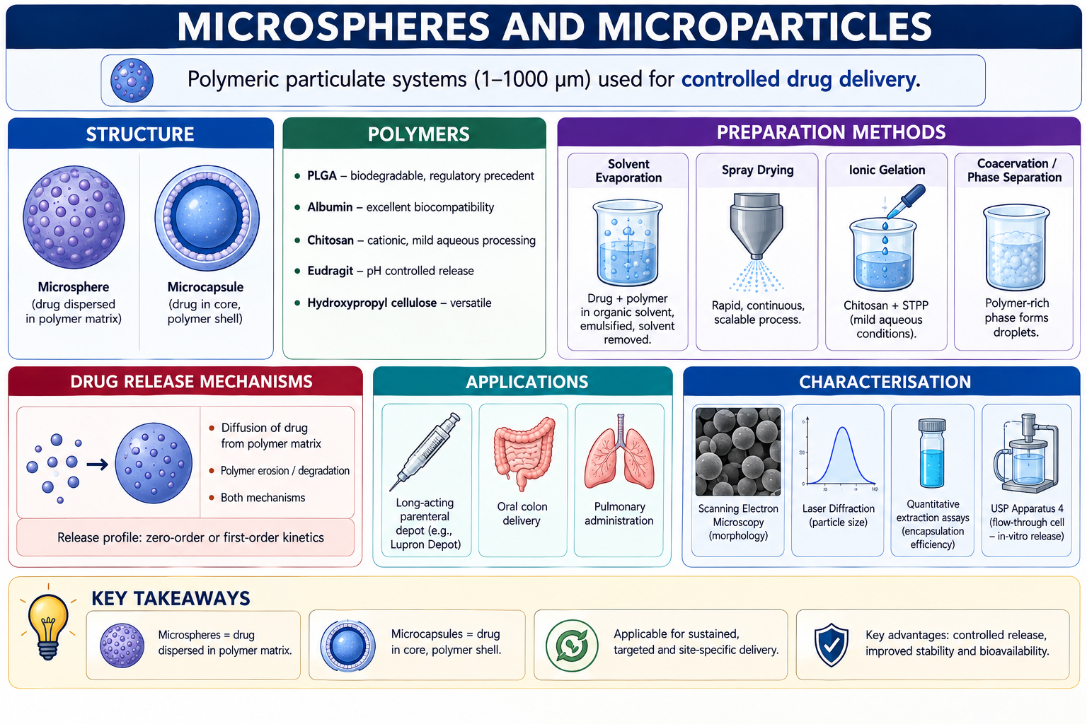
Microspheres and microcapsules are polymeric particulate systems, typically ranging from one to one thousand micrometres in diameter, distinguished from one another by their internal architecture: a microsphere consists of a homogeneous polymeric matrix throughout which the drug is dispersed or dissolved, whereas a microcapsule possesses a distinct core-shell structure, with the drug confined within a reservoir enclosed by a discrete polymeric membrane. The polymers most frequently employed for microparticle fabrication include PLGA, valued for its established biodegradability and regulatory precedent; albumin, an endogenous protein offering excellent biocompatibility; chitosan, a cationic polysaccharide amenable to mild, aqueous-based ionic gelation processing; and various grades of Eudragit and hydroxypropyl cellulose, selected according to the desired release profile and administration route.
Microparticle preparation methods are selected according to the physicochemical properties of the drug and the desired release characteristics, and include solvent evaporation, in which drug and polymer dissolved in a volatile organic solvent are emulsified into an aqueous continuous phase and the solvent subsequently removed to harden the resulting droplets into solid microspheres; spray drying, a rapid, continuous, and readily scalable process well suited to industrial manufacture; ionic gelation, particularly applicable to chitosan systems through crosslinking with sodium tripolyphosphate under mild aqueous conditions favourable to labile biologics; and coacervation or phase separation techniques.
Drug release from microparticulate systems proceeds through diffusion of drug from the polymer matrix, through erosion or degradation of the polymer itself, or through a combination of both mechanisms operating simultaneously, and the resulting release profile may approximate zero-order or first-order kinetics depending on the dominant mechanism and the specific polymer and formulation employed. Clinically, microspheres have achieved particular commercial success as long-acting parenteral depot formulations, exemplified by the PLGA-based product Lupron Depot, which provides sustained release of a peptide hormone over periods extending to several months from a single injection, as well as in applications targeting oral colon delivery and pulmonary administration. Characterisation of microparticulate systems relies on scanning electron microscopy for morphological assessment, laser diffraction for particle size determination, quantitative extraction assays for encapsulation efficiency, and in-vitro release testing, frequently employing USP Apparatus 4 flow-through cell methodology, to establish the release kinetics of the finished product.
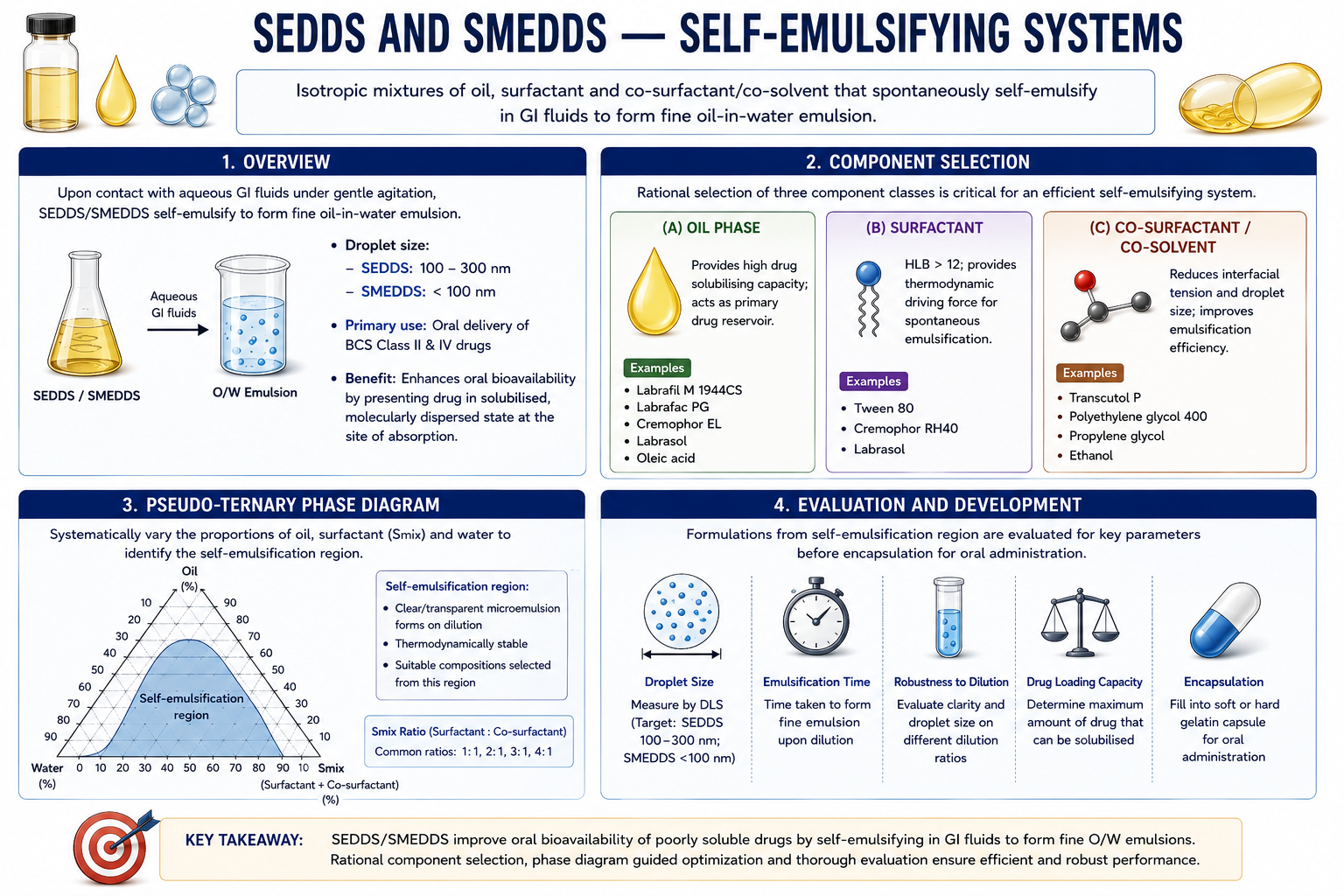
Self-Emulsifying Drug Delivery Systems, abbreviated SEDDS, and their finer-droplet variant, Self-Microemulsifying Drug Delivery Systems, abbreviated SMEDDS, are isotropic mixtures of oil, surfactant, and co-surfactant or co-solvent that, upon exposure to aqueous gastrointestinal fluids under the gentle agitation provided by normal gut motility, spontaneously self-emulsify to form a fine oil-in-water emulsion, with resulting droplet sizes in the range of 100 to 300 nanometres for conventional SEDDS and below 100 nanometres for the more finely dispersed SMEDDS. These systems find their primary pharmaceutical application in the oral delivery of BCS Class II and Class IV drugs, where they can produce dramatic improvements in oral bioavailability by presenting the drug already in a solubilised, molecularly dispersed state at the point of intestinal absorption, circumventing the dissolution step that otherwise rate-limits absorption for poorly soluble compounds.
The rational design of a SEDDS formulation proceeds through the systematic screening of three component classes. Oil phase selection prioritises components offering high drug solubilising capacity, such as Labrafil M 1944CS, Labrafac PG, Cremophor EL, Labrasol, or oleic acid, since the oil serves as the primary drug reservoir within the formulation. Surfactant selection favours agents with a Hydrophile-Lipophile Balance value exceeding twelve, such as Tween 80, Cremophor RH40, or Labrasol, since these hydrophilic surfactants provide the principal thermodynamic driving force for spontaneous emulsification upon aqueous dilution. Co-surfactants or co-solvents, including Transcutol P, polyethylene glycol 400, propylene glycol, or ethanol, are incorporated to further reduce interfacial tension and droplet size, enhancing the overall efficiency and speed of the self-emulsification process.
Optimisation of SEDDS and SMEDDS formulations is conventionally guided by construction of a pseudo-ternary phase diagram, in which the relative proportions of oil, surfactant, and aqueous phase are systematically varied and the resulting phase behaviour recorded, allowing identification of the self-emulsification region — the zone of composition space within which spontaneous, clear microemulsion formation occurs upon aqueous dilution. Formulations selected from within this self-emulsification region are subsequently evaluated for droplet size, emulsification time, robustness to dilution, and drug loading capacity, before progressing to encapsulation, typically within a soft or hard gelatin capsule shell, for oral administration.
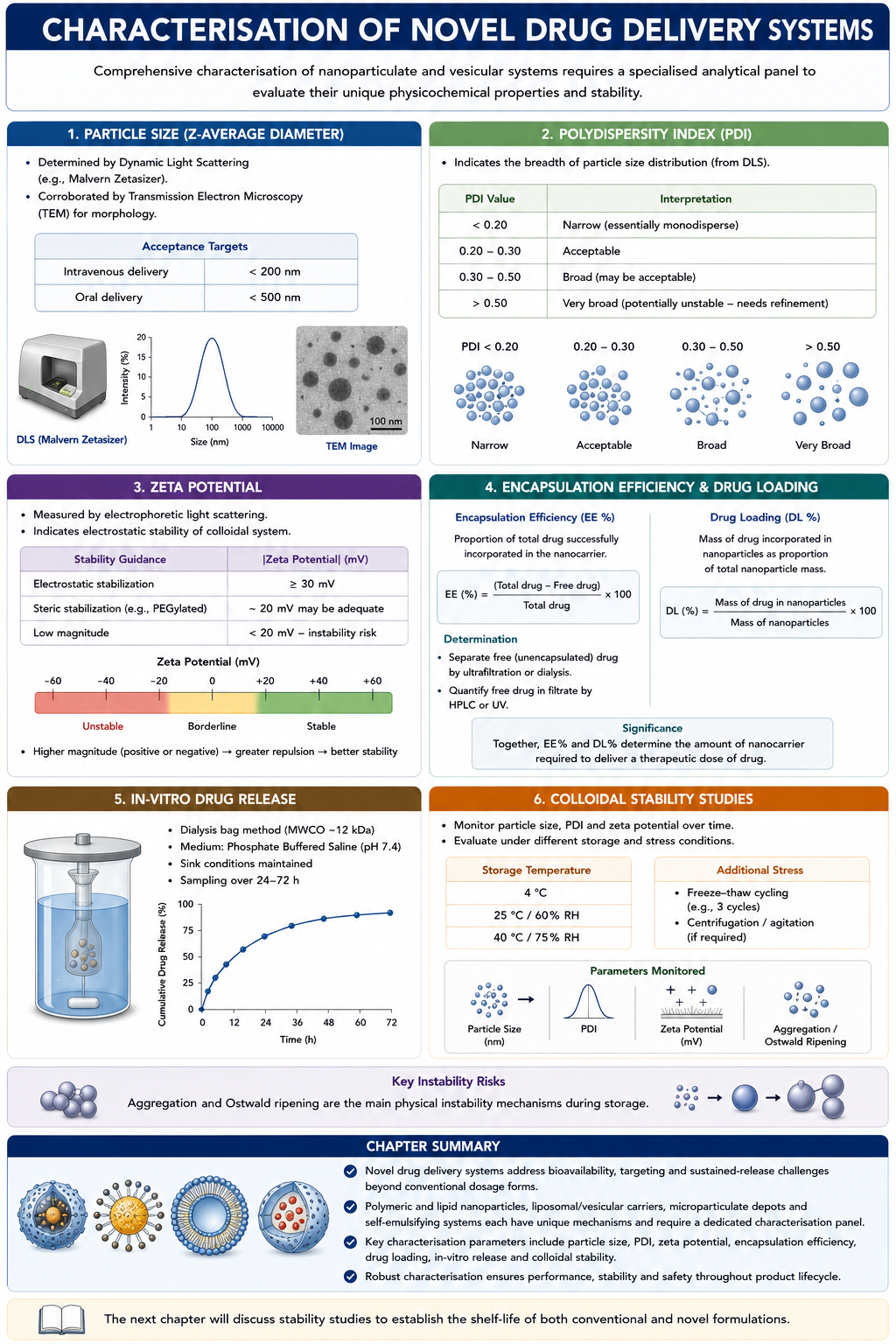
The comprehensive characterisation of nanoparticulate and vesicular delivery systems requires a distinct analytical panel beyond that applied to conventional dosage forms, reflecting the unique physical properties and stability considerations of colloidal systems. Particle size, expressed as the Z-average diameter, is most commonly determined by Dynamic Light Scattering using instruments such as the Malvern Zetasizer, with acceptance targets generally below 200 nanometres for intravenous administration and below 500 nanometres for oral delivery, and findings are frequently corroborated by Transmission Electron Microscopy to provide direct morphological confirmation. The Polydispersity Index, also derived from Dynamic Light Scattering, quantifies the breadth of the particle size distribution, with values below 0.2 indicating a narrow, essentially monodisperse population, values up to approximately 0.3 generally regarded as acceptable, and values exceeding 0.5 indicative of a broad, potentially unstable distribution requiring formulation refinement.
Zeta potential, a measure of the electrostatic charge at the particle surface derived from electrophoretic mobility measurements, provides a key indicator of colloidal stability, since particles bearing a sufficiently strong surface charge, conventionally at least thirty millivolts in magnitude, resist aggregation through electrostatic repulsion, while particles stabilised primarily through steric hindrance, for instance by a PEGylated surface coating, may remain colloidally stable at somewhat lower zeta potential magnitudes, around twenty millivolts, owing to the additional steric contribution to stability.
Encapsulation Efficiency, conventionally abbreviated EE per cent, quantifies the proportion of total drug successfully incorporated within the nanocarrier, calculated as the difference between total and free drug content divided by total drug content, and is typically determined indirectly by separating unencapsulated free drug through ultrafiltration or dialysis followed by high-performance liquid chromatography or ultraviolet quantification of the drug remaining in the filtrate. Drug Loading, a related but distinct parameter, expresses the mass of drug incorporated as a proportion of the total nanoparticle mass, and together these two parameters determine the practical dose of nanocarrier material a patient must receive to achieve a given therapeutic dose of drug.
In-vitro release from nanoparticulate systems is typically assessed using a dialysis bag method, employing a membrane with a molecular weight cut-off around twelve kilodaltons immersed in phosphate-buffered saline at pH 7.4 under sink conditions, with sampling extended over twenty-four to seventy-two hours to capture the often prolonged release kinetics characteristic of these systems. Finally, colloidal stability is assessed by monitoring particle size, polydispersity index, and zeta potential over one to three months at a range of storage temperatures, typically 4, 25, and 40 degrees Celsius, together with resistance to freeze-thaw cycling, since aggregation and Ostwald ripening represent the principal physical instability risks confronting nanoparticulate formulations during long-term storage.